Jurkat Cells 96 Well Plate
Super Resolution Imaging Of Jurkat T Cells In A 96 Well Plate With Download Scientific Diagram

Attachment Of Jurkat Cells Was Assayed In 96 Well High Binding Plates Download Scientific Diagram

Jurkat Cells T Cell Leukemia T Cell Signaling Molecular Devices
A Simple Technique For Quantifying Apoptosis In 96 Well Plates Bmc Biotechnology Full Text

Effect Of Acacetin On The Expression Of Kv1 3 Channels In Jurkat Cells Download Scientific Diagram

Cell Meter Phosphatidylserine Apoptosis Assay Kit Red Fluorescence Optimized For Microplate Readers Aat Bioquest
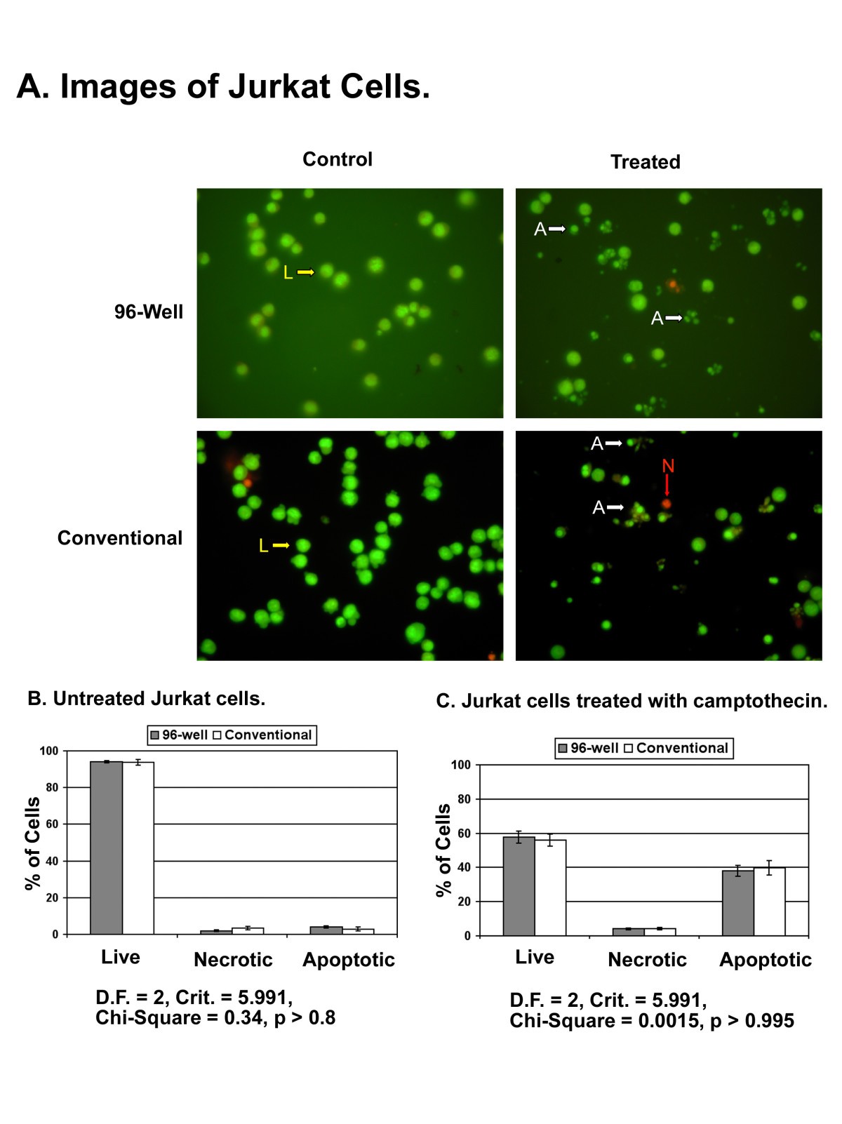
Eb ao dye mix 8 μl was added to each well and cells were viewed under the same microscope as above.
Jurkat cells 96 well plate. Stainfree cell counts vs. 50 000 and 1562 cells seeded per well of a 96 well plate. 1 reduction of the time period during the test and the processing of the results. For both suspension and adherent cells 96 well plates were centrifuged at 1 000 rpm 129 g for 5 minutes using a beckman model tj 6 centrifuge with inserts for 96 well plates.
Prepare aliquots to avoid repeated freeze thaw cycles. Jurkat zellen die bei zelldichten von 390 bis 50 000 zellen pro well ausgesät wurden wurden mit der stainfree technologie blaue punkte analysiert oder sie wurden mit dem farbstoff des earlytox live cell assays gefärbt und die grün fluoreszierenden zellen ausgezählt grüne punkte. Incubate at 37 c for 2 hours or 4 c overnight. Tests were done in triplicate counting a minimum of 100 total cells each.
3 several conditions can be tested at the same time. Plate 200 μl of cell culture i e 50 000 200 000 cells into the wells of the sterile 96 well filter bottom plate. Back to the gibco cell culture basics homepage. For this table hela cells were used.
Fluorescent cell counts jurkat cells seeded at densities ranging from 390 to 50 000 cells per well were counted using stainfree technology blue dots or they were stained with earlytox live cell assay dye and green fluorescent cells were counted green dots. I haven t worked so much with jurkat cells so far but for the experiements we made we seeded in a 96 well plate with 50 000 200 000 cells. Storage and stability store quanti luc pouches at 20 c for 12 months. Stimulate the cells as desired.
When α cd3 coating is done wash 6 well plate 1 x 5 ml pbs then add cells to each well do not plate more than 5 ml well minimum is 2 ml. For cd4 t cells incubation is usually done at. I guess you can adjust the number by just trying it out. Die mit beiden methoden bestimmten zellzahlen stimmen über alle zelldichten hinweg nahezu überein.
For the unstimulated control wells add 50 µl of sterile pbs. Wash plate microwells 3 times with sterile pbs. This new adhesion technique has several advantages in comparison with its predecessor. Two cell densities are shown.
Aseptically decant antibody solution from the microwell plate. Dispense 50µl of the antibody solution to each microwell of the 96 well assay plate. 1 the number of cells on a confluent plate dish or flask will vary with cell type. Therefore we have developed a new method performed in a 96 well plate format adding the quantification of the non adherent viable cells with a colorimetric reaction.
4 quantification of viable. Each pouch contains everything needed to prepare 25 ml of reagent allowing the preparation of 500 wells of a 96 well plate. The filter plate is designed to retain particles while permitting the flow of liquids from the bottom of the plate. 2 the experiments can be done in a larger scale.
In the example presented below jurkat cells were stimulated with 20 μg ml anisomycin for 60 minutes at 37 c.

A High Throughput Method For Characterizing Novel Chimeric Antigen Receptors In Jurkat Cells Sciencedirect

536i05

Attachment Of Jurkat Cells Was Assayed In 96 Well High Binding Plates Download Scientific Diagram
Ijms Free Full Text Physiological Responses Of Jurkat Lymphocytes To Simulated Microgravity Conditions Html
Https Www Activemotif Com Documents 1539 Pdf

Amplite Fluorimetric Caspase 3 7 Assay Kit Red Fluorescence Aat Bioquest
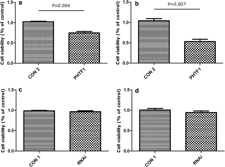
Analysis Of The Expression Of Phtf1 And Related Genes In Acute Lymphoblastic Leukemia Cancer Cell International Full Text
Biomolecules Free Full Text Effects Of Fludioxonil On The Cell Growth And Apoptosis In T And B Lymphocytes Html

A Assay Process Involving Jurkat T Cell Stimulation And Tacrolimus Download Scientific Diagram

Signosis
Genome Editing Isolating Clones For Genotypic And Phenotypic Characterization

Cross Linking Of Ephb6 Resulting In Signal Transduction And Apoptosis In Jurkat Cells The Journal Of Immunology

Cell Meter Multiplexing Caspase 3 7 8 And 9 Activity Assay Kit Triple Fluorescence Colors Aat Bioquest